Sunday, March 30, 2014

X-ray angiography directly to good effect in breast cancer screening

X-ray contrast media is directly the best way to check for breast cancer,has become popular for use in home and abroad. 
Breast (x-ray machine)X-ray angiography is the best means of breast cancer screening and early diagnosis.Canadian researchers report that on the 14th,there are two digital breast X-ray angiography for use in direct digital radiography (DR) screening is better than a computer X -ray contrast (CR). 
Angiography difference between the two is that similar to a digital camera directly to angiography,the radiologist can instantly via computer image analysis;while the computer angiography breast image information is recorded in a contrast panel,read by the reading device,the computer processing an image is formed. 
mobile x-ray machine 
Ontario,Canada"Cancer Care Ontario"project researchers in the American Journal of the 14th issue of"Radiology"the report said,breast cancer screening in Ontario in addition to two digital (portable x-ray machine)X-ray angiography,as well as the use of traditional film X line angiography.810,000 times their breast cancer screening from 2008 to 2009 in the province of more than 680,000 women were surveyed.It was found that direct digital radiography and conventional film angiography screening effect is quite,but the detection rate of breast cancer is relatively low computer angiography. 
Typically,every 1000 women receiving breast (mobile x-ray machine)X-ray angiography screening will find less than five cases of breast cancer.Study reported that the use of computers angiography,per 10,000 women screened will be less about 10 cases of breast cancer screening. 
The article comes from:http://www.medicalequipment-msl.com/htm/medical-device-book/X-ray-angiography-directly-to-good-effect-in-breast-cancer-screening.html 
Related Posts 
The first American X-ray 
China's analog X-ray machines are gobbling up US film supply

Thursday, March 27, 2014

How to quickly raise the level of ultrasound diagnostic methods and techniques

1.master a variety of skills.Skills are the basic conditions for wrist ultrasound diagnosis.Good operating practices,image display is clear,it becomes relatively easy to diagnose.As"look good,play good not strong."I often take less time in patients in patients with operating practices Duan Lian,over time,potentially increasing their operating practices.Baoding Fifth Hospital Division seized power on behalf of Yahui. 
2.solid and extensive clinical knowledge to improve the diagnosis premise.Diagnostic ultrasound is a clinical purposes,an ultrasound image can be a variety of diseases ultrasonography.Thus,ultrasound can not plug-speak,we must combine with the clinical phase. 
bovine ultrasound machines 
3.strengthen expertise in bovine ultrasound machine diagnosis of learning is essential to improve the conditions.As an ultrasound workers should have the solid foundation of professional knowledge. 
4.focusing on the positioning-Qualitative-Training Differential Diagnosis way of thinking.Positioning is the key,is to guide diagnosis.Some of lesion localization is very easy to very simple,but sometimes it is difficult,such as in the liver,kidney,and the junction of the right adrenal gland disease,positioning is quite easy.Qualitative is perfect,is the key to diagnosis.Differential diagnosis is corroborated.Established diagnosis is inseparable from the differential diagnosis.Let the differential diagnosis of a reliable basis for diagnosis,but with real results even closer match. 
5.the follow-up.Follow-up ultrasound machines for sale diagnosis for improving the level of play for a very important role.Every time we finished a complicated person from the disease,the diagnosis is often made,but my heart still want to know the final outcome of surgery and pathology.Follow-up is the only way to follow-up allows us to make a correct diagnosis of the disease sonogram further deepen understanding of missed diagnosis and misdiagnosis for patients looking for the wrong reasons which,to avoid the view that after the corresponding error,so as to achieve the purpose of improving the diagnostic level. 
The article comes from:http://www.medicalequipment-msl.com/htm/medical-device-book/How-to-quickly-raise-the-level-of-ultrasound-diagnostic-methods-and-techniques.html 
Related Posts 
How Ultrasound Works 
Ultrasonography in patients with low back pain lumbar artery blood flow detection application

Wednesday, March 26, 2014

How Long Are Premature Babies Kept in Incubators?

There is no set amount of time for a premature baby to stay in an incubator. Hospitals place most very premature babies or sick babies on open warming beds shortly after birth, so that it is easier to assess them and work on them. Once the baby needs fewer interventions, he generally goes into an incubator, which is an enclosed space that keeps him warm. Once he can maintain his temperature, he will go into an open crib -- the final step before coming home. How long the process takes depends on your baby. 
Baby incubator 
Temperature Stability 
The key to making the move from incubator to open crib is your baby's ability to maintain his temperature on his own in room air. Because premature babies do not have as much brown adipose tissue, a type of baby fat or as much subcutaneous fat as full-term babies do, they have more difficulty maintaining their temperature without a heated environment. Premature infants also experience insensible water loss from their increased surface area; keeping them in an incubator reduces fluid loss. 
Weight 
Your baby's weight helps determine when he is ready to try a move to an open crib. Many hospitals consider that a baby is able to try an open crib once he reaches 1,700 to 1,800 grams, or between 3 pounds, 12 ounces to 3 pounds, 15 ounces, according to an Australian study published in the 2011 "Cochrane Neonatal Reviews." After a review of available studies, the researchers concluded that hospital staff can safely move premature infants to an open crib when they weigh at 1,600 grams, or 3 pounds, 8 ounces -- or possibly, even less. 
Gestational Age 
Babies start to produce brown adipose tissue, or BAT, between the gestational age of 26 to 30 weeks, according to Birth.com. By 40 weeks, healthy newborns have enough BAT to maintain their temperatures in normal indoor temperatures without difficulty. Sometime between these two points, your baby will be ready for an open crib, but his current weight and medical conditions will determine when he is ready. 
Other Factors 
Infections, illness and stress can affect your preemie's ability to maintain his temperature adequately. Because of this, he might need to remain in an Cheap Isolettes even though he weighs as much as a normal full-term baby. A baby who cannot maintain his temperature might not gain weight as well as a baby kept in an incubator, because he uses up additional energy keeping his temperature stable. 
The article comes from:http://www.medicalequipment-msl.com/htm/medical-device-book/How-Long-Are-Premature-Babies-Kept-in-Incubators.html

Monday, March 24, 2014

Ultrasonography in patients with low back pain lumbar artery blood flow detection application

bovine ultrasound machines plays an important role in the examination of the lumbar spine pain.Especially in the lumbar artery exploration. 
Maintain adequate blood supply due to the normal function of the lumbar spine is very important:for capillary blood from the bone between the lumbar artery;disc of the student movement from the bone between the capillary network;destroy these blood vessels and blood flow can lead to disc blood for other important structural barriers to yield the corresponding disease state.Some scholars believe that the abdominal aorta of atherosclerotic plaques and other diseases can cause arterial transportation waist reduced,thereby inducing disc degeneration and low back pain,the study of vascular hemodynamic waist help clinicians better understand them and disc degeneration,low back pain and other related properties. 
color ultrasound machine 
Currently waist vascular hemodynamic data on the normal population,and the population is still not reported back pain,and color Doppler ultrasound as a non-invasive examination methods assisted hemodynamic monitoring,which is currently in clinical applications have been increasingly the more extensive,and its hemodynamic measurements have confirmed the reliability of data for clinical research institute.Recently,scholars from Oxford University orthopedic applications on color Doppler ultrasound in the detection of blood flow dynamics in patients with low back pain,a technical report,and compare the normal population and in patients with low back pain associated hemodynamic data and conclusions published in Spine magazine. 
Study is divided into two groups,one group with low back pain,low back pain for three months,in the past by MRI,patients diagnosed with lumbar disc degeneration presence of 64 patients ( 35 females and 29 males,age range 21 - 82 years;mean age of 50 ± 16.5 years);normal group,back pain does not occur within the previous June had not received medical treatment because of back pain,a total of 30 cases (13 females and 17 males,age range 19 - 82 years,mean age 41 ± 18.5 years). 
Radiographic evaluation methods:carry blood from the rear of the lumbar spine to detect.Start L5-S1 begin to determine the fifth lumbar artery and sacral artery,then up probe to determine 4th - 1st lumbar artery ( Figure 1 ). 
Figure 1:Color Doppler ultrasound probe(color ultrasound machine),more visible vessel branches emanating from the extension of the lumbar facet joints spinous 
Get bilateral lumbar artery hemodynamic data,the use of Doppler ultrasound-assisted software records the peak systolic velocity (peak systolic velocity,PSV,Figure 2 ).A total of 12 data,each side 6.Changes produced in order to avoid the situation of human physiological impact on the statistical data,the PSV values standardized calculation PSVR value (PSV ratio)=lumbar artery PSV values aortic PSV values. 
Figure 2:Color Doppler ultrasound showed the traffic branch. 
The results showed that : 
The normal population,lumbar artery PSV values,showing a gradual increasing trend from the first to the fourth lumbar arteries lumbar artery,while the smallest in the fifth lumbar artery PSV values;PSV values sacral artery has increased over the fifth lumbar artery,but Less than a fourth lumbar artery;there were significant differences (p <0.001) between the segmental arteries compare PSV,but about bilateral comparison was no significant difference (p=0.09),as shown in Table 1. 
Table 1:lumbar artery PSV 
Aortic PSV shown in Table 2,the probe found no evidence of aortic atherosclerosis;although PSV's statistical difference (p <0.01) exists between the low back pain group and the normal group,but these differences are not of clinical significance because the two groups of patients with aortic PSV were within the normal range. 
Table 2:PSV value aorta 
Low back pain group and normal group PSVR comparison:Table 3 shows a comparison of the data back pain group and the normal group PSVR data area.Two aspects of the same trend increase in PSVR:L1-L4 increases,L5 reduced to a minimum value,S1 rise again,there is a significant difference (p <0.001) between segments comparison;between back pain group segments PSVR value than the normal population set high (p <0.001),between groups and segments no significant correlation values (p=0.9).Table 4 shows the PSV 95% confidence interval,Figure 3 shows the various groups PSVR values on different segments. 
Table 3:PSVR value distribution 
Table 4:PSV 95% confidence interval 
Figure 3:Each group PSVR values on different segments of 
Conclusions:PSVR value of low back pain patients was significantly higher than the normal population,the characteristics and Doppler ultrasound vascular congestion(cheap ultrasound machine) when the change is similar to changing conditions and dynamics of the relationship between the pathogenesis of low back pain in patients with low back pain blood still needs further clarification. 
The article comes from:http://www.medicalequipment-msl.com/htm/medical-device-news/Ultrasonography-in-patients-with-low-back-pain-lumbar-artery-blood.html 
Related Posts 
How Ultrasound Works 
Ultrasound use in the frozen process

Friday, March 21, 2014

Surgical shadowless eight advantages-Part II.


Fifth,easy to operate


During surgery requires frequent changes in position and direction of the operation of the lamp according to the surgery,the operation must be simple,flexible,lightweight,brake quickly and effectively.Should be set to adjust the focus,illumination devices.Beaten the United States and other countries produce surgical lights,light body central portion with an operating handle can be sterilized(mini autoclave sterilizer),the surgeon can always adjust the position and direction,focus and illumination lamps.
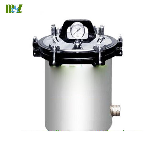
Portable stainless steel sterilizer-MSLPS02

Six,with good response performance


During surgery experience a sudden power outage,affecting the smooth operation,the consequences are self-evident,qualified medical units are fitted with captive power generation and lighting,to prepare for emergency use.As the ideal surgical lights should have AC-DC composite supply,power supply automatic switching devices.Can better cope with a sudden power outage,always protect the operation can proceed smoothly.1970s,the United States,Japan,Germany and other federal army has good performance of field equipment emergency surgical lights.In 1984,the People's Liberation Army Institute of Medical Equipment made of high illumination,low temperature,AC-DC composite supply,the power automatically switched AC voltage regulator.DC power supply for a long time,the new structure of the 84-type multi-function mobile shadowless.Such surgery lighting equipment can not only meet usually use as well shadowless illumination performance and comfort,but also have a high degree of mobility and wartime use function in harsh environments. 

Seven,economic security is better


Economy is mainly refers to the high light-emitting efficiency,low power consumption,long life.Active use of new technologies,new processes,new materials(bench top autoclave).Efforts to improve product quality and reduce costs.Large foreign halogen bulbs and light sources has been adopted by repeatedly refracted light cold light projection equipment.When you use halogen bulbs,attention should be not more than offset the positive and negative supply voltages of 2.5%.This is because the supply voltage varies greatly on the lamp life and light effects influence;When the voltage is increased 5%,the life will be shortened by 50%.Recently the structure of large and complex surgical lights tend to consider their safety is very important,it mainly refers to the electrical safety performance.Electrical installations and wiring connections shall comply without installing devices to ensure the absolute safety of use. 

Eight others


Theater is one of the best medical facilities in the area clean.Light body should step on the adhesion of dust made it difficult material,smooth surface,there should be no bump phenomenon in order to keep the lamp body clean sweep.Surgical lights structural strength and weight can not be ignored,as far as possible using high quality lightweight materials.Major surgery light weight does not exceed 100 kg.Note that when mounted hanging mechanical security.
In recent years,with the rapid development of medical career,in order to meet the needs of modern surgery,surgical lights for the increasingly high demand at home and abroad have been developed to produce high illumination,low power consumption,long life new surgical lights.As senior product containing fiber deep Designed,color(color ultrasound machine) television photographic equipment and ventilation devices.In order to meet the needs of the surgeon set brightness adjustment device on high intensity surgical lights.Angle of illumination by light energy needs surgery and surgical scope changes.Buried want surgical lights should provide adequate illumination,good light color effect,groups of high,low temperature,high-quality and comfortable uniform beam.Taking into account the conditions in other areas to meet the needs of modern surgery.
The article comes from:http://www.medicalequipment-msl.com/htm/medical-device-book/Surgical-shadowless-eight-advantages-Part-II.html
Related Posts
Eight surgical shadowless advantage-Part I

Tuesday, March 18, 2014

Eight surgical shadowless advantage-on the articles.


Is a very important surgical shadowless lighting operation.It is used for special operations in the lighting,the surgical safety,quality of surgery,surgical efficiency and the ability to continue to work for the surgeon,eye health has a significant impact.With the development of the cause of medical care,surgical shadowless proposed new requirements.As ideal surgical shadowless should meet the following conditions(bovine ultrasound machine). 

A good shadowless effect


Usually surgical lamps called surgical light.This is a few dozen or a ring light evenly ball 50 to 100 cm in diameter in the shade device.Players using a special light bulb and reflector,the light source gathered from various angles in a subject according to the surface.This object is illuminated under shadowless encountered staggered interaction of light,reflected in the shadow of irradiated surface becomes extremely slight ghost,get a clear surgical field during surgery.As surgical shadowless lamp must have a good effect.Shadowless degree is an important feature and performance indicators surgical lamp.Any shadows in the surgical field will hinder the formation of the surgeon's observation,identification and surgery.Ideal surgical lamp in addition to providing adequate illumination,shall shadowless must have a high degree of assurance the surgical field surface and organizations have a deep luminosity.Part of the operation shadowless lamp see diagram.
Eight surgical shadowless 

Second,according to high


Surgery is the most intense visual task.Task lighting should be high illumination to reduce visual fatigue and improve work efficiency.About fine job lighting standards,national regulations vary.Depending on the object to another is less than 0.15 mm in diameter,for example,France is 1200 lux,Germany 2000 lux,3000 lux Britain,the United States 5000 to 10,000 lux,Japan 700 to 1500 lux,lux country is tentatively scheduled for 2000.Different surgical site,and some deep body cavity surgery,requires concentrating well,according to high,according to the Japanese Industrial Standard JISZ9110~1969,the provisions in the surgical field diameter of 30 cm range illumination of 2000 lux.High intensity surgical lights must have the condition.Recently,the Japanese developed a surgical light illumination of up to 2,000,000 lux.As surgical lighting,most countries believe that to 60,000 to 100,000 lux illumination is appropriate,the equivalent of direct sunlight at noon in summer outdoor illumination is too bright can affect vision.Provide adequate illumination at the same time,to avoid glare on the beam surgical instruments.Glare affect vision and visual,easy to make eye fatigue,is not conducive to the smooth progress of surgery.Illumination and hand surgery room 's overall illumination lamp should be a certain percentage of the illumination should not differ between the two Mrs.America provisions in the operating room illumination than 1000 lux.Japan provided the overall lighting illumination is 1:10 local lighting illumination,that the overall illumination of the operating room should be above 1000 lux. 

Third,the low temperature heat less


With the improvement of illumination,and improve the beam will cause the temperature of the lamp body.Direct thermal beam head surgeon and surgical site tissue surgical lights will affect the efficiency and quality of operation surgery.To this end,the world attach great importance to this issue,and constantly develop new surgical lights,efforts aimed at reducing the temperature of the lamp.Recently,the French developed into a theater with a new cold light projection lamp.The lamp comprises an optical system and a number of cold mirrors.Using this light,compared with all previous surgery lighting equipment.Produce a stronger light,and heat was small.Due to the characteristics of the cold mirror,the incident light is only visible light region,the absolute output of the lamp is cold output.Up to 135,000 lux of illumination by changing the size and the focus position of the illumination area.In recent years,the use of chemical drugs,alternating layers of plating method on complex reflector(automatic x ray film processor) to heat and condenser.Again in front of the lamp assembly a heat wave reflection coated film complex devices.Visible light generated by the light source may be filtered through the infrared reflector 70%,most of the remaining infrared isolate and filter glass.After the above process called optical Cambodia cold.This cold surgical lights,heat not only small and high illumination,is an ideal surgical lighting equipment.Being finalized publication"People's Republic of China national military standard GJB-87 field lights," clearly stipulates the surgical field temperature:Run field surgical lights should not exceed 4C; portable field surgical lights should not exceed 6C. 

Fourth,good color effect light


The so-called surgical lighting color effect,provided that the beam can truly reflect the current blood(hematology analyzer),tissue and change the color.Same color object under light irradiation with different spectral power distributions show different colors.Color light show illuminated object color performance is called the light source.Surgical light required,not only to see the color of blood,and must be able to clearly distinguish the change of color and changes in blood tissue.Requires surgical lamp light source with excellent color rendering.Under incandescent seen with blood seen under a fluorescent color is different.Nature of the object is determined by the color of the color of the light source.Provisions of the international reference source CRI of 100.High color rendering index,color distortion less,that color is a good source;while the lower CRI,the more serious distortion,color of the light source that is not good.For example: incandescent bulbs and halogen tube general color rendering index of 95 to 99 ),with fluorescent tubes general color rendering index of 65-80).Color rendering and color temperature of the light source are closely related.Object emits a high temperature heat and light conditions,and changing the light color temperature.Light color available color temperature,the unit is the absolute temperature K.6000K color temperature required to achieve the true color of about 70 to 90,the effective color is close to 100.
The article comes from:http://www.medicalequipment-msl.com/htm/medical-device-book/Eight-surgical-shadowless-advantage-on-the-articles.html