Since the discovery of X-rays, two-dimensional (2-D) radiographic imaging has been the most widely used approach for ultrasound imaging anatomic structures, diagnosing disease, and guiding interventions . Unfortunately, 2-D X-ray imaging cannot provide physicians with a complete set of information fo imaging the human body.Although 3-D CT, MRI, and X-ray techniques(x ray protection) are important imaging modalities, 3-D ultrasound machine has been shown to play a major role in medical diagnostics and minimally invasive image-guided interventions.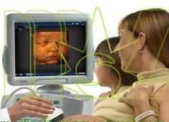 The basic concept of 3-D ultrasound is to collect a data of 2-D ultrasound images (black &white of color ) while tracking and storing the location of each individual 2-D image . The data set is then reconstructed into a single 3-D volume that can be displayed on the screen and manipulated. The reconstructed 3-D volume can be rotated, sliced, rendered or displayed in multiplanar cross-sections. Three-dimensional ultrasound imaging has provided high-definition images of complex anatomic structures and pathology to diagnose disease and to monitor and guide interventional procedures.Although 2-D arrays are available for real-time 3-D imaging, most low-cost 3-D ultrasound(portable ultrasound machine) imaging systems use conventional one-dimensional (1-D) ultrasound transducers to acquire a series of 2-D US images. These images are reconstructed to form a 3-D image. A wide variety of approaches have been developed to determine the position and orientation of the 2-D images within the 3-D image volume. The production of a 3-D ultrasound image volume, without any distortions, requires that three factors be optimized:The scanning technique must be either rapid (e. g., real time) or gated to avoid image artifacts due to involuntary, respiratory, or cardiac motion.The locations and orientations of the acquired 2-D ultrasound image must be accurately known to avoid geometric distortions in the 3-D ultrasound image, which could lead to errors of measurement and guidance.The scanning apparatus must be simple and convenient to use, so that the scanning process can be easily added to an examination or interventional procedure.Because a set of 2-D ultrasound images is used to reconstruct the 3-D ultrasound image, the transducer must be moved so that the set of acquired 2-D ultrasound images covers the region of interest. However, a 2-D array allows the transducer to remain stationary , an electronic scanner is then used to sweep the ultrasound bean over the entire volume, allowing for the acquisition of a set of 3-D images to occur in real time (i.e. , 4-D ultrasound imaging ).In this approach , a 2-D phased array of transducer elements is used to transmit a broadly diverging beam of ultrasound away from the array, sweeping out a volume shaped like a truncated pyramid. The returned echoes detected by the 2-D array are processed to display a set of multiple plane in real time . Using various image rendering techniques, user can interactively control and manipulate these planes to explore the entire volume. This approach is successfully used in echocardiography.Many algorithms have been developed to help physicians and researchers visualize and manipulate 3-D medical images interactively. Because ultrasound image suffer from image speckle and poor tissue-tissue contrast, the display of a 3-D image plays a dominant role in the ability of physician to obtain valuable information.Moreover, Doppler ultrasound technology also be developed, for example. Spectral Doppler scanning displays information about the spectrum of flow velocities as a function of time. It is sometimes called FFI (Fast Fourier Transform) because the information is presented as a frequency spectrum indicating velocity components.The article comes from:http://www.medicalequipment-msl.com/htm/medical-device-news/ultrasound-imaging-technology.htmlFor more information about medical equipments visit our website.
Since the discovery of X-rays, two-dimensional (2-D) radiographic imaging has been the most widely used approach for ultrasound imaging anatomic structures, diagnosing disease, and guiding interventions . Unfortunately, 2-D X-ray imaging cannot provide physicians with a complete set of information fo imaging the human body.Although 3-D CT, MRI, and X-ray techniques(x ray protection) are important imaging modalities, 3-D ultrasound machine has been shown to play a major role in medical diagnostics and minimally invasive image-guided interventions.The basic concept of 3-D ultrasound is to collect a data of 2-D ultrasound images (black &white of color ) while tracking and storing the location of each individual 2-D image . The data set is then reconstructed into a single 3-D volume that can be displayed on the screen and manipulated. The reconstructed 3-D volume can be rotated, sliced, rendered or displayed in multiplanar cross-sections. Three-dimensional ultrasound imaging has provided high-definition images of complex anatomic structures and pathology to diagnose disease and to monitor and guide interventional procedures.Although 2-D arrays are available for real-time 3-D imaging, most low-cost 3-D ultrasound(portable ultrasound machine) imaging systems use conventional one-dimensional (1-D) ultrasound transducers to acquire a series of 2-D US images. These images are reconstructed to form a 3-D image. A wide variety of approaches have been developed to determine the position and orientation of the 2-D images within the 3-D image volume. The production of a 3-D ultrasound image volume, without any distortions, requires that three factors be optimized:The scanning technique must be either rapid (e. g., real time) or gated to avoid image artifacts due to involuntary, respiratory, or cardiac motion.The locations and orientations of the acquired 2-D ultrasound image must be accurately known to avoid geometric distortions in the 3-D ultrasound image, which could lead to errors of measurement and guidance.The scanning apparatus must be simple and convenient to use, so that the scanning process can be easily added to an examination or interventional procedure.Because a set of 2-D ultrasound images is used to reconstruct the 3-D ultrasound image, the transducer must be moved so that the set of acquired 2-D ultrasound images covers the region of interest. However, a 2-D array allows the transducer to remain stationary , an electronic scanner is then used to sweep the ultrasound bean over the entire volume, allowing for the acquisition of a set of 3-D images to occur in real time (i.e. , 4-D ultrasound imaging ).In this approach , a 2-D phased array of transducer elements is used to transmit a broadly diverging beam of ultrasound away from the array, sweeping out a volume shaped like a truncated pyramid. The returned echoes detected by the 2-D array are processed to display a set of multiple plane in real time . Using various image rendering techniques, user can interactively control and manipulate these planes to explore the entire volume. This approach is successfully used in echocardiography.Many algorithms have been developed to help physicians and researchers visualize and manipulate 3-D medical images interactively. Because ultrasound image suffer from image speckle and poor tissue-tissue contrast, the display of a 3-D image plays a dominant role in the ability of physician to obtain valuable information.Moreover, Doppler ultrasound technology also be developed, for example. Spectral Doppler scanning displays information about the spectrum of flow velocities as a function of time. It is sometimes called FFI (Fast Fourier Transform) because the information is presented as a frequency spectrum indicating velocity components.The article comes from:http://www.medicalequipment-msl.com/htm/medical-device-news/ultrasound-imaging-technology.htmlFor more information about medical equipments visit our website.
Sound generated above the human hearing range is called ultrasound(cheap ultrasound machine). Although ultrasound behaves in a similar manner to audible sound, it has a much shorter wave-length. This means it can be reflected off very small surfaces such as defects inside materials. It is this property that makes ultrasound useful for medical testing and medical measure. Ultrasonic vibrations travel in the form of a wave, similar to the way light travels. However, unlike light waves which can travel in a vacuum (empty space), ultrasound requires an elastic medium such as liquid or a solid. Ultrasonic nondestructive testing introduces high frequency sound waves into a test object to obtain information about the object without altering or damaging it in any way. Two basic quantities are measured in ultrasonic testing; they are the time of flight or the amount of time for the sound to travel through the sample and the amplitude of received signal. Based on velocity and round trip time of flight through the material, the material thickness can be calculated. 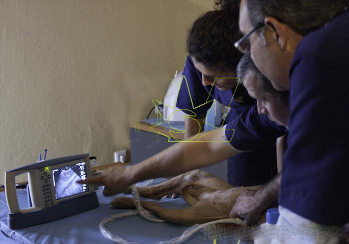 Measurements of the relative change in signal amplitude can be used in sizing flaws or measuring the attenuation of a material. The relative change in signal amplitude is commonly measured in decibels. Decibel values are the logarithmic value of the ratio of two signal amplitudes.General operation of each block is described below.
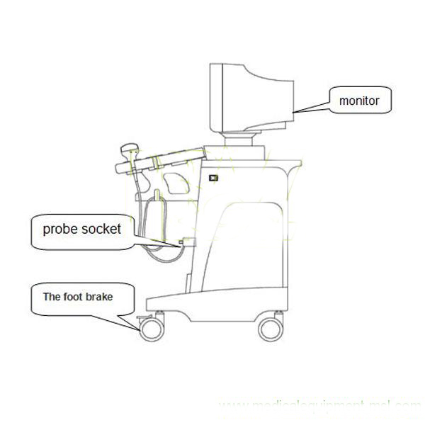 Transmitter & ReceiverThis block provides the burst wave by receiving the control signal transmitted from DSC (digital scan converter). It generates the transmission trigger TRIG 1 to n, based on this burst wave TRIG 1 to n are send to probe. Received echo 1 to n sent from probe are amplified under the control of GAIN and STC (sensitivity time compensation), and sent to DSC as echo video signal.Probe The probe has some blocks of circuits consisting of the transmitter amp, receiver preamp, multi-channel analogue multiplexer and counter. TRIG 1 to n sent from Transmitter & Receiver is amplified by the transmitting amp, so as to generate the pulse for driving the transducer. The echo signal received by the transducer is amplified by the preamplifier and sent to Transmitter & Receiver Board. Analogue multiplexers are connected to 64 elements of transducers and perform the electronic scan by the control signal. A transducer is any device that converts one form of energy to another. An ultrasonic transducer converts electrical energy to mechanical energy in the form of sound, and vice versa.
 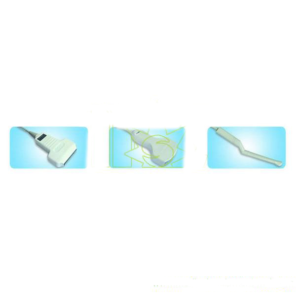 DSUltrasound echo video signal sent from Transmitter & Receiver is converted to 4-bit digital data by A/D converted video data of 8 pixels are stored and they are written in image Memory at the same time. Memory address for writing are generated synchronizing with Ultrasound transmission timing. Memory address for writing are generated synchronizing with TV-scanning signal. Data of 8 pixels are also read out at the same time and using parallel-serial conversion, Data of each pixel can be obtained. Post-process by line-interpolation is performed by adding Data read out from Image Memory and Data read out from Line Memory. Ultrasound image is smoothed by this processing method. Digital ultrasonic(medical equipments) data are added with graphic data read out Graphic Memory, gray-scale data and composite sync signal sent from Control PCB, and then sent to D/A converter to be converted to analogue TV signal (composite video). Cleaning on Picture TubeAs Picture Tube is always apt to adsorb dusts in air because static electricity rise and therefore the resolution would be lowered, please keep it clean periodically. Caution on CheckingIt is necessary for you to take care on checking, because transistor is very weak in electric shock and would be broken when voltage over its rated value. As especially Horizontal Circuit is of and because a minor mistake in handling sometimes cause transistor to break, please do not check high voltage caused by means of making sparks with a screw driver, etc. The article comes from:http://www.medicalequipment-msl.com/htm/medical-device-book/ultrasonic-apparatus.html For more information about aluminum crutches price visit our website.
Sound generated above the human hearing range is called ultrasound(cheap ultrasound machine). Although ultrasound behaves in a similar manner to audible sound, it has a much shorter wave-length. This means it can be reflected off very small surfaces such as defects inside materials. It is this property that makes ultrasound useful for medical testing and medical measure. Ultrasonic vibrations travel in the form of a wave, similar to the way light travels. However, unlike light waves which can travel in a vacuum (empty space), ultrasound requires an elastic medium such as liquid or a solid. Ultrasonic nondestructive testing introduces high frequency sound waves into a test object to obtain information about the object without altering or damaging it in any way. Two basic quantities are measured in ultrasonic testing; they are the time of flight or the amount of time for the sound to travel through the sample and the amplitude of received signal. Based on velocity and round trip time of flight through the material, the material thickness can be calculated. Measurements of the relative change in signal amplitude can be used in sizing flaws or measuring the attenuation of a material. The relative change in signal amplitude is commonly measured in decibels. Decibel values are the logarithmic value of the ratio of two signal amplitudes.General operation of each block is described below. Transmitter & ReceiverThis block provides the burst wave by receiving the control signal transmitted from DSC (digital scan converter). It generates the transmission trigger TRIG 1 to n, based on this burst wave TRIG 1 to n are send to probe. Received echo 1 to n sent from probe are amplified under the control of GAIN and STC (sensitivity time compensation), and sent to DSC as echo video signal.Probe The probe has some blocks of circuits consisting of the transmitter amp, receiver preamp, multi-channel analogue multiplexer and counter. TRIG 1 to n sent from Transmitter & Receiver is amplified by the transmitting amp, so as to generate the pulse for driving the transducer. The echo signal received by the transducer is amplified by the preamplifier and sent to Transmitter & Receiver Board. Analogue multiplexers are connected to 64 elements of transducers and perform the electronic scan by the control signal. A transducer is any device that converts one form of energy to another. An ultrasonic transducer converts electrical energy to mechanical energy in the form of sound, and vice versa. DSUltrasound echo video signal sent from Transmitter & Receiver is converted to 4-bit digital data by A/D converted video data of 8 pixels are stored and they are written in image Memory at the same time. Memory address for writing are generated synchronizing with Ultrasound transmission timing. Memory address for writing are generated synchronizing with TV-scanning signal. Data of 8 pixels are also read out at the same time and using parallel-serial conversion, Data of each pixel can be obtained. Post-process by line-interpolation is performed by adding Data read out from Image Memory and Data read out from Line Memory. Ultrasound image is smoothed by this processing method. Digital ultrasonic(medical equipments) data are added with graphic data read out Graphic Memory, gray-scale data and composite sync signal sent from Control PCB, and then sent to D/A converter to be converted to analogue TV signal (composite video). Cleaning on Picture TubeAs Picture Tube is always apt to adsorb dusts in air because static electricity rise and therefore the resolution would be lowered, please keep it clean periodically. Caution on CheckingIt is necessary for you to take care on checking, because transistor is very weak in electric shock and would be broken when voltage over its rated value. As especially Horizontal Circuit is of and because a minor mistake in handling sometimes cause transistor to break, please do not check high voltage caused by means of making sparks with a screw driver, etc. The article comes from:http://www.medicalequipment-msl.com/htm/medical-device-book/ultrasonic-apparatus.html For more information about aluminum crutches price visit our website.
Eupes Inc., a diagnostic ultrasound company, is an innovator in medical devices(x ray protection) for acute care and private practice health care providers.Since it’s Aug.8, 1988 founding in a modest basement workshop, Eupes has grown to become an international firm, with the company’s products now used by health care providers in over 80 countries. With offices in the U.S., Canada, the UK, China, France, and Australia, the company emloys over 360 people worldwide.Gary R. Smith, an electrical engineer, founded the company, with the mission to offter meaningful improvement in patient care to the health care community. Eupes, recognized as a leader in both portable ultrasound(cheap ultrasound machine) and the revolutionary field of laryngoscopy, was ranked among the fastest growing technology companies in Massachusetts in Deloitte’s prestigious ”Technology Fast 50” program in 2001, 2002, 2005, 2007 and 2009. Gary Smith was named winner of the Ernst & Young Entrepreneur of the Year Award in the Northeast for the Health services category in 2009. And the company was acknowledged as one of the best places to work in Massachusetts by Massachusetts CEO Magazine, in both 2006 and 2009.Eupes is best known for its BladderSee brand, a line of bladder volume measurement in struments that has fuled the growth of the company from a small start-up to a profitable international copporation. Noninvasive and easy to use, BladderSee is clinically proven to prevent unnecessary urinary catheterizations and reduce rates of urinary tract infection (UTI). This is of significant importance to hospitals, in light of changing Medicare reimbursement for catheter-related UTIs, beginning in October, 2008. BladderSee also helps diagnose urinary-related problems such as en-larged prostate (BPH) and bladder outlet obstruction (BOO), of importance to the growing number of Baby Boomers and the health care providers who treat them. Now a standard of care for noninvasive bladder volume measurement, the BladderSee is used in Urology, Acute Care, Pri-mary Care and Extended Care.The company expanded its product portfolio in January 2007 with the acquisition of Asped Biomedical Systems and the GlideView brand. GlideView Laryngoscope provides a consistently clear view of the airway, enabling quick intubation, and offer significant benefits to Anesthesiolo-segment of NBC’’s”ER-The Final Season.” The GlideView device is currently being used by the military in both Afghanistan and Iraq.The article comes from:http://www.medicalequipment-msl.com/htm/medical-device-book/Eupes-GlideView.htmlFor more information about portable ultrasound machine visit our website.
Eupes Inc., a diagnostic ultrasound company, is an innovator in medical devices(x ray protection) for acute care and private practice health care providers.Since it’s Aug.8, 1988 founding in a modest basement workshop, Eupes has grown to become an international firm, with the company’s products now used by health care providers in over 80 countries. With offices in the U.S., Canada, the UK, China, France, and Australia, the company emloys over 360 people worldwide.Gary R. Smith, an electrical engineer, founded the company, with the mission to offter meaningful improvement in patient care to the health care community. Eupes, recognized as a leader in both portable ultrasound(cheap ultrasound machine) and the revolutionary field of laryngoscopy, was ranked among the fastest growing technology companies in Massachusetts in Deloitte’s prestigious ”Technology Fast 50” program in 2001, 2002, 2005, 2007 and 2009. Gary Smith was named winner of the Ernst & Young Entrepreneur of the Year Award in the Northeast for the Health services category in 2009. And the company was acknowledged as one of the best places to work in Massachusetts by Massachusetts CEO Magazine, in both 2006 and 2009.Eupes is best known for its BladderSee brand, a line of bladder volume measurement in struments that has fuled the growth of the company from a small start-up to a profitable international copporation. Noninvasive and easy to use, BladderSee is clinically proven to prevent unnecessary urinary catheterizations and reduce rates of urinary tract infection (UTI). This is of significant importance to hospitals, in light of changing Medicare reimbursement for catheter-related UTIs, beginning in October, 2008. BladderSee also helps diagnose urinary-related problems such as en-larged prostate (BPH) and bladder outlet obstruction (BOO), of importance to the growing number of Baby Boomers and the health care providers who treat them. Now a standard of care for noninvasive bladder volume measurement, the BladderSee is used in Urology, Acute Care, Pri-mary Care and Extended Care.The company expanded its product portfolio in January 2007 with the acquisition of Asped Biomedical Systems and the GlideView brand. GlideView Laryngoscope provides a consistently clear view of the airway, enabling quick intubation, and offer significant benefits to Anesthesiolo-segment of NBC’’s”ER-The Final Season.” The GlideView device is currently being used by the military in both Afghanistan and Iraq.The article comes from:http://www.medicalequipment-msl.com/htm/medical-device-book/Eupes-GlideView.htmlFor more information about portable ultrasound machine visit our website.
Singapore’s medical device(elbow crutches for sale) market is expected to grow as the island state strengthens its reputation as the region’s healthcare hub and center of healthcare excellence. Demand for state of the art medical technologies is high as Singapore strives to provide first class healthcare delivery systems and facilities to its residents as well as serve the international patient market.The U.S., Japan and Germany are the top three leading suppliers of medical equipment in Singapore. There are significant number of companies that have established a presence by setting up their regional headquarters there in an effort to be closer to and better serve their customers. The U.S. enjoys a good reputation and is recognized by the industry as technologically superior, providing high quality, advanced and reliable equipment. However, the U.S. is not price competitive in the lower-end consumables, typically supplied by manufacturers in more labor-abundant countries throughout Asia.Singapore has a mixed healthcare market comprising competent public and private providers and together they provide excellent healthcare services and offer choices to both Singaporeans and foreign patients.Demand for medical devices comes from public and private hospitals and clinics. The Ministry of Health (MOH) is the largest consumer, accounting for nearly 70% of local demand. The Parkway Group, owner of the three largest private hospitals in Singapore, is also a significant consumer of medical devices.Price and quality are often cited as primary factors that determine purchasing decisions of medical devices. Other considerations are reliable and prompt after-sales service. For large government procurement, purchases are typically made via tender announcements.There are central purchasing departments consisting of a team from the various public sector hospital’s Material Management Departments (MMDs) who work together to achieve economies of scale for large purchases.Typically, hospitals make their own purchasing decisions and source for products through the MMDs in the various hospitals. They will in turn consult with the end-users of the medical equipment on the product specifications.There are no custom duties on medical devices. A 5.0% goods and services tax (GST) is imposed on all goods sold and services provided, locally. Imports are subject to GST, but payments are refundable on re-exports.Foreign companies who are new to the market and interested in exporting to Singapore pay consider appointing a local distributor to represent their company’s products and services. Given the small market size of the island state, most potential distributors would request for exclusive rights to sell the product. This will ensure that they commit their resources to promoting the product to the appropriate end-users and reap the returns should a sale materialize.Depending on the medical equipment, the foreign company will be required to either provide samples, or accord special rates to the potential distributor for “demo units”. The potential distributor will use the samples to conduct a survey of the market to ascertain interest in the product(aluminum crutches price) while the “demo units” will be used as they demonstrate the products’ technology to the potential buyers.As the sales in the local market increases, the foreign company can look into setting up an on-going presence in Singapore much like how some large MNCs have set up regional offices in Singapore. This brings the foreign company closer to their customers, demonstrates their commitment to the region and allows for prompt and enhanced customer service.Foreign companies can take part in regional trade shows, international medical device fairs, or sector-specific conferences where they can showcase their products, technologies and services.The article comes from:medical device
Singapore’s medical device(elbow crutches for sale) market is expected to grow as the island state strengthens its reputation as the region’s healthcare hub and center of healthcare excellence. Demand for state of the art medical technologies is high as Singapore strives to provide first class healthcare delivery systems and facilities to its residents as well as serve the international patient market.The U.S., Japan and Germany are the top three leading suppliers of medical equipment in Singapore. There are significant number of companies that have established a presence by setting up their regional headquarters there in an effort to be closer to and better serve their customers. The U.S. enjoys a good reputation and is recognized by the industry as technologically superior, providing high quality, advanced and reliable equipment. However, the U.S. is not price competitive in the lower-end consumables, typically supplied by manufacturers in more labor-abundant countries throughout Asia.Singapore has a mixed healthcare market comprising competent public and private providers and together they provide excellent healthcare services and offer choices to both Singaporeans and foreign patients.Demand for medical devices comes from public and private hospitals and clinics. The Ministry of Health (MOH) is the largest consumer, accounting for nearly 70% of local demand. The Parkway Group, owner of the three largest private hospitals in Singapore, is also a significant consumer of medical devices.Price and quality are often cited as primary factors that determine purchasing decisions of medical devices. Other considerations are reliable and prompt after-sales service. For large government procurement, purchases are typically made via tender announcements.There are central purchasing departments consisting of a team from the various public sector hospital’s Material Management Departments (MMDs) who work together to achieve economies of scale for large purchases.Typically, hospitals make their own purchasing decisions and source for products through the MMDs in the various hospitals. They will in turn consult with the end-users of the medical equipment on the product specifications.There are no custom duties on medical devices. A 5.0% goods and services tax (GST) is imposed on all goods sold and services provided, locally. Imports are subject to GST, but payments are refundable on re-exports.Foreign companies who are new to the market and interested in exporting to Singapore pay consider appointing a local distributor to represent their company’s products and services. Given the small market size of the island state, most potential distributors would request for exclusive rights to sell the product. This will ensure that they commit their resources to promoting the product to the appropriate end-users and reap the returns should a sale materialize.Depending on the medical equipment, the foreign company will be required to either provide samples, or accord special rates to the potential distributor for “demo units”. The potential distributor will use the samples to conduct a survey of the market to ascertain interest in the product(aluminum crutches price) while the “demo units” will be used as they demonstrate the products’ technology to the potential buyers.As the sales in the local market increases, the foreign company can look into setting up an on-going presence in Singapore much like how some large MNCs have set up regional offices in Singapore. This brings the foreign company closer to their customers, demonstrates their commitment to the region and allows for prompt and enhanced customer service.Foreign companies can take part in regional trade shows, international medical device fairs, or sector-specific conferences where they can showcase their products, technologies and services.The article comes from:medical device
FDA of USA is cautioning of malware that can breach the security of medical devices, which have configurable embedded PCs, published infosecurity-magazine.com, June 17, 2013.FDA alerts that since there's a growing interconnection of medical devices through the Internet, smart-phones, hospital networks alternatively more medical devices, cyber-security infiltrations into medical devices(cheap medical equipment) are increasing, posing danger to the operations of such equipments. Initiation of the assaults is possible through the use of malicious software against the devices alternatively, via illegal admission into hospital networks' or medical devices' configuration setups, the agency warns.In 2012 autumn, IOActive realized a fact that a laptop could be used to remotely regulate as well as instruct many suppliers' pacemakers for delivering shock as intense as 830 volt, which was actually possible due to flawed software programs that configured the medical devices. Such efforts are certainly sufficient to kill a user, while Barnaby Jack Company Researcher observed that the security flaws provided opportunity for 'bulk killing.'Disturbingly, FDA's alert is not just an announcement for, problem cases have been spotted occurring all across the Web.And though it's not in the knowledge of FDA regarding death/injury of any patients because of the above kind of incidents, nor is there any sign to it about any particular computer or equipment in clinical application being deliberately targeted, still the agency decided to proceed in this aspect in close collaboration with manufacturers as well as other federal associations for the detection, communication and minimization of security flaws and consequent attacks.In its advisory, the FDA gives suggestion to both health-care providers and device makers.It wrote that expectedly, medical device makers would adopt suitable measures for restricting the situations that allowed illegal admission into medical equipments. Precisely, the device makers required examining their online security policies and practices for maintaining suitable protections from illegal admission into their products else allowance of modifications to the same just as they should defend against cyber-security hijacking of the device-connected hospital network, it explained. Softpedia.com published this, June 17, 2013.To health-care providers, FDA suggested they should prevent illegal admission into their networks as well as interconnected medical equipments.The article comes from:http://www.medicalblogvoice.com/cyber-hacking-into-medical-devices-possible-cautions-fda.html